The Histology of Brittle Nail Syndrome (Onychoschizia)
Fingernails are specialized epithelial appendages composed of approximately 80 to 90 layers of compact, flattened onychocytes welded together by intercellular membrane complexes and hard alpha-keratin fibers. Brittle nail syndrome (onychoschizia) is characterized by impaired intercellular adhesion between dorsal and intermediate nail lamellae, leading to horizontal transverse splitting, distal fraying, and vertical ridging.
Frequent exposure to water, household cleaning solvents, and alcohol sanitizers leaches natural lipids and bound water out of the nail plate, making it mechanically fragile. However, topical nail polishes and cuticle oils only coat the superficial dead plate; they cannot alter the structural architecture of new nail tissue. Healing onychoschizia requires delivering systemic nutrients to the generative nail matrix buried beneath the proximal cuticle.
Targeted Nutritional Recovery with High-Dose Biotin & Zinc
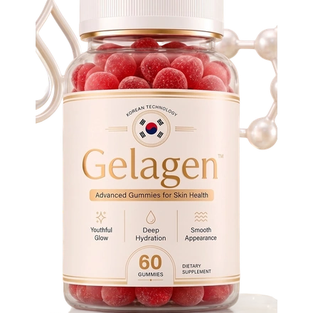
In clinical trials by Colombo and Scher, subjects with documented brittle nails received daily high-dose biotin therapy. Scanning electron microscopy confirmed that within three to six months, new nail plates emerging from the proximal fold displayed a 25% increase in thickness, smooth surface morphology, and dramatic resistance against transverse splitting. Gelagen combines 5000 mcg D-biotin with Zinc Citrate and Ascorbic Acid to maximize onychocyte keratin cross-linking from within.